

With the help of 3-D printers, even hobbyists can make model rockets or gaming accessories out of seemingly nothing. What if that printer could make new body parts, such as an ear, lung or skin? This may sound like science fiction. But scientists are already using living cells to “ink” prototypes of these tissues.
This 3-D bioprinting uses the same basic strategy now used to print toys, sculptures, metal pieces or even basketballs. Specialized printers just swap out plastic or metal for living cells.
Explainer: What is 3-D printing?
Actually, there’s a bit more to it than that. In most cases, you can’t use the same devices to make tissues. And you can’t just spew cells onto a surface, layer by layer, and expect them to link up into working organs. The cells have to be mixed with some soft, gel-like materials to make them unite with their neighbors.
Although still experimental, some devices have already printed skin, cartilage and even early-stage blood vessels. In 2025, researchers in Australia used an experimental robot to bioprint ink made from a patient’s own skin cells directly onto her burn wounds.
One of the doctors who pioneered the bioprinting of skin onto burn wounds describes how it’s done and why her team is excited about it.It starts with the ink
In most 3-D printers, a resin, plastic filament or metal serves as the ink. Bioprinters instead use cells — and something to keep them alive.
“Bioinks are specialized soft gels that can have living cells mixed inside them,” explains Ryan Martin. He works on this tech at Virginia Commonwealth University, or VCU, in Richmond. Think of these inks like Jell-O: wobbly, soft materials that can hold their shape while protecting what’s inside.
Explainer: What is a hydrogel?
Scientists make these hydrogels from materials such as alginate (AL-jin-ate) and collagen. Alginate comes from seaweed. Collagen is a protein found in skin and bones.
But bioinks contain more than just cells and hydrogel. That extra stuff — part of a complex matrix — helps the mix mimic the material that surrounds cells in the body, explains Ankita Pramanick. She’s a biomedical engineer at Utrecht University in the Netherlands.
Bioprinted tissue in space
 NASA Johnson Space Center/Wikimedia Commons
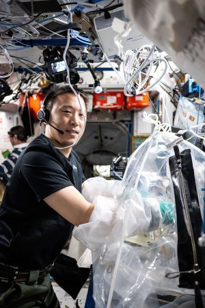
NASA astronaut Jonny Kim treats bioprinted liver tissue on September 17, 2025, in a portable glove-bag on the International Space Station. These samples later went into another research device so researchers could study the effects of microgravity on blood vessel formation in engineered tissues.
What makes bioinks different from most other 3-D inks is that they host living cells. And those cells must survive the printing process, where they’ll get squeezed through nozzles and exposed to temperature changes. The matrix is designed with these challenges in mind.
After printing, the inked structures go into incubators, which are containers that maintain ideal conditions to help cells grow. There, the newly printed cells can mature, strengthen and grow together to form tissues — perhaps cartilage for joints or layered skin with blood vessels.
Printing living cells
Engineers have been developing several different ways to turn bioinks into tissue. Which one they choose depends on what they’re building.
Most times, a “bioink is pushed through a nozzle like toothpaste from a tube,” Martin explains. This process is known as extrusion. A machine controls how fast the ink comes out, building up a tissue layer by layer.
For example, to create skin tissue, you’d first print fibroblasts (FY-broh-blasts) — the cells that make up the inner layer of your skin. Later, you’d layer on keratinocytes (Kare-uh-TIN-oh-sites). These are the cells that make up our skin’s protective outer layer. Once printed, this combined structure matures within an incubator.
Extrusion works well for thicker, structured tissues. These include skin and cartilage. But it’s relatively slow. Squeezing cells through a nozzle can also stress them.
Inkjet bioprinting is faster and gentler. It sprays tiny droplets of bioink onto a surface in much the same way an office printer sprays ink onto paper. It works best with a very liquid matrix. That’s why it’s often used for thin cell layers, such as those that line the insides of blood vessels.
Another type of bioprinting creates entire structures at once instead of building them layer by layer. To start, a laser shines its light into a spinning tube of light-sensitive gel and cells. This solidifies them into the precise shape needed.
With this, scientists might print a small network of blood vessels all at once — complete with all its branches. It could take just seconds. And that’s important, because speed matters. The faster tissues are printed, the less time their cells spend outside their ideal environment.
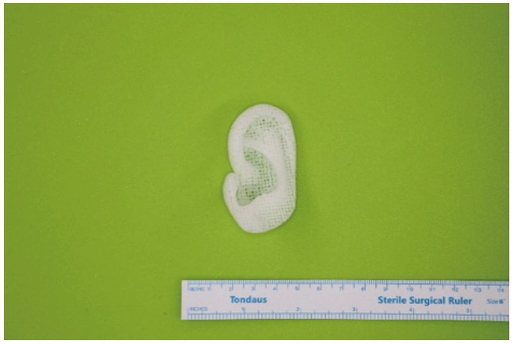 This 3-D printed ear implant was made from polycaprolactone (PCL), a biodegradable polymer. The implant is custom-designed to match a patient’s own ear and is shown next to a ruler for scale. The goal is to transition from plastic, as here, to implants made from living cells.M. Kim et al./Yonsei Medical Journal 2024 (CC BY-NC 4.0)
What’s being printed
In 2022, a patient received a 3-D–printed ear made from their own cells. In late 2025, surgeons successfully implanted a bioprinted cornea — the clear outer layer of the eye — into someone who had been legally blind.
Skin grafting is perhaps the most advanced application. A skin graft is a type of transplant where healthy skin is used to cover missing or injured skin. A 2025 trial in Australia tested bioprinted skin on burn patients. In a news conference with her doctor, the first patient to receive such a graft showed it had successfully worked on her leg.
Bone is trickier to print. Unlike skin or cartilage, it contains hard minerals mixed with living cells. Scientists have used 3-D printing to create bone scaffolds — frameworks that encourage the body’s own bone cells to grow and fill in the gaps. But no one has yet bioprinted actual bone with living cells in it for people.
Complex, fully-functioning organs such as hearts and kidneys are still a work in progress. Tissues thicker than a few millimeters (about a tenth of an inch) face a critical problem: Shortly after printing, their cells quickly start dying from a lack of oxygen and nutrients.
Heart of the matter
 A Pramanick et al./Advanced Functional Materials 2025 (CC BY 4.0)
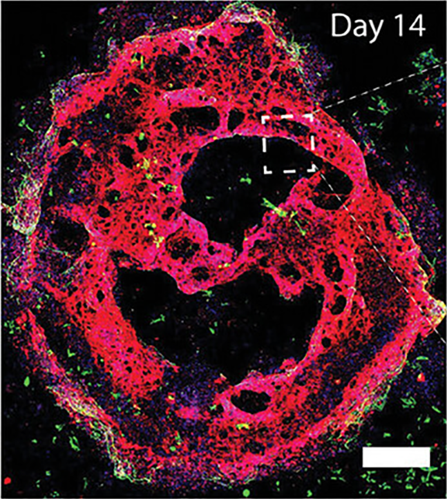
The image shows bioprinted heartlike tissue after 14 days in an incubator. The different colors help show the position of different types of cells inside the tissue.
The missing piece: blood vessels. These are needed for bringing oxygen to cells deep inside thick tissues and carrying away wastes. Without that oxygen and waste removal, cells will die. That’s why building blood vessels into tissues is the biggest hurdle standing in the way of fully printed organs.
The problem comes down to diffusion — the movement of molecules from areas of high concentrations to low concentrations. Oxygen can diffuse only a few millimeters into tissue. In thicker tissues, cells in the center would suffocate before oxygen could reach them. Imagine a big, densely populated city with no roads. Food trucks can’t bring in supplies. Garbage trucks can’t haul away waste.
Scientists have tried ways to avoid this problem. One method uses sacrificial inks. These serve as temporary placeholders. The inks are printed in the shape of vessels, then tissue is printed around them. Once the tissue solidifies, the sacrificial ink dissolves. In its place sit hollow channels through which blood can flow.
Scientists are also looking for a way to print blood vessels directly into tissue as it’s being built up.
Do you have a science question? We can help!
Submit your question here, and we might answer it an upcoming issue of Science News Explores
Moving forward
This short overview describes what bioprinting is, how advanced it’s become and where it might go.In September 2025, researchers made a promising advance with a 3-D bioprinting system they’re calling GRACE. ”This technology enables the printing of blood vessels directly around [the printed tissue itself],” explains Pramanick at Utrecht. She works in the lab that’s developing GRACE.
Here’s how it works. A computer-vision system “sees” where cells are located. It then asks artificial intelligence to figure out the best pattern of blood vessels to deliver oxygen-rich blood to all those cells. In seconds, bioprinting then creates pipe-shaped structures to begin delivering that blood.
Speed matters enormously here. Traditional methods might take hours to print vessel networks layer by layer. Because GRACE does it in seconds, cells spend little time without oxygen.
Full organs remain years away. Forming stable, treelike structures in different sizes — from big arteries down to the tiniest vessels known as capillaries — to seamlessly work with a patient’s blood-vessel system remains a big challenge, notes Martin at VCU. As bioinks and printing speeds improve, the prospect of integrating printed tissues with blood vessels into the body appears ever nearer. Today, the question is no longer if 3-D bioprinting can create replacement organs — it’s when.



 Bengali (Bangladesh) ·
  English (United States) ·