

Last year, scientists achieved a historic first by providing a patient with the first-ever corneal implant made solely of human cells grown in the lab.
Corneal transplants are used to treat severe corneal scarring and inflammation, eye injuries and complications from eye surgeries. They replace the clear dome at the front of the eye, and usually, the tissues for the procedure are collected from organ donors after death. But in this case, one donor's corneal tissue was used to create hundreds of implants through sophisticated laboratory culture techniques and 3D bioprinting.
This technology, created by Precise Bio, an Israeli regenerative medicine company also based in North Carolina, produces a transparent, layered structure that resembles a healthy, natural cornea. The approach could help to reduce the scarcity of donor corneas worldwide, the company says. An early-stage trial, known as a Phase I trial, is currently underway to evaluate the technique's safety in human patients.
To learn more about the science behind the 3D-printed corneas and the next steps for development, Live Science spoke with Precise Bio co-founders Aryeh Batt and Dr. Anthony Atala.
Neelanjana Rai: How are most corneas for transplants sourced and prepared, currently?
Anthony Atala: They are typically recovered from deceased donors within a few hours of death and then banked. They go through screening and testing, and are then preserved until implanted — usually within a couple of weeks — before being transplanted to patients in need.
NR: Are there problems or limitations with this approach?
AA: Absolutely. In fact, availability is a major challenge. There is a very limited donor supply and a significant worldwide shortage of transplantable corneas, meaning a lot of patients lack access to corneas for implantation, which is a huge deficit.
Aryeh Batt: Today, for every cornea transplant performed worldwide, roughly 70 people remain without one because there are not enough donor corneas. There are between 12 million to 15 million people worldwide in need of a cornea transplant who do not have access to donor tissue.
AA: The ultimate goal is to completely eliminate the shortage of these corneas.
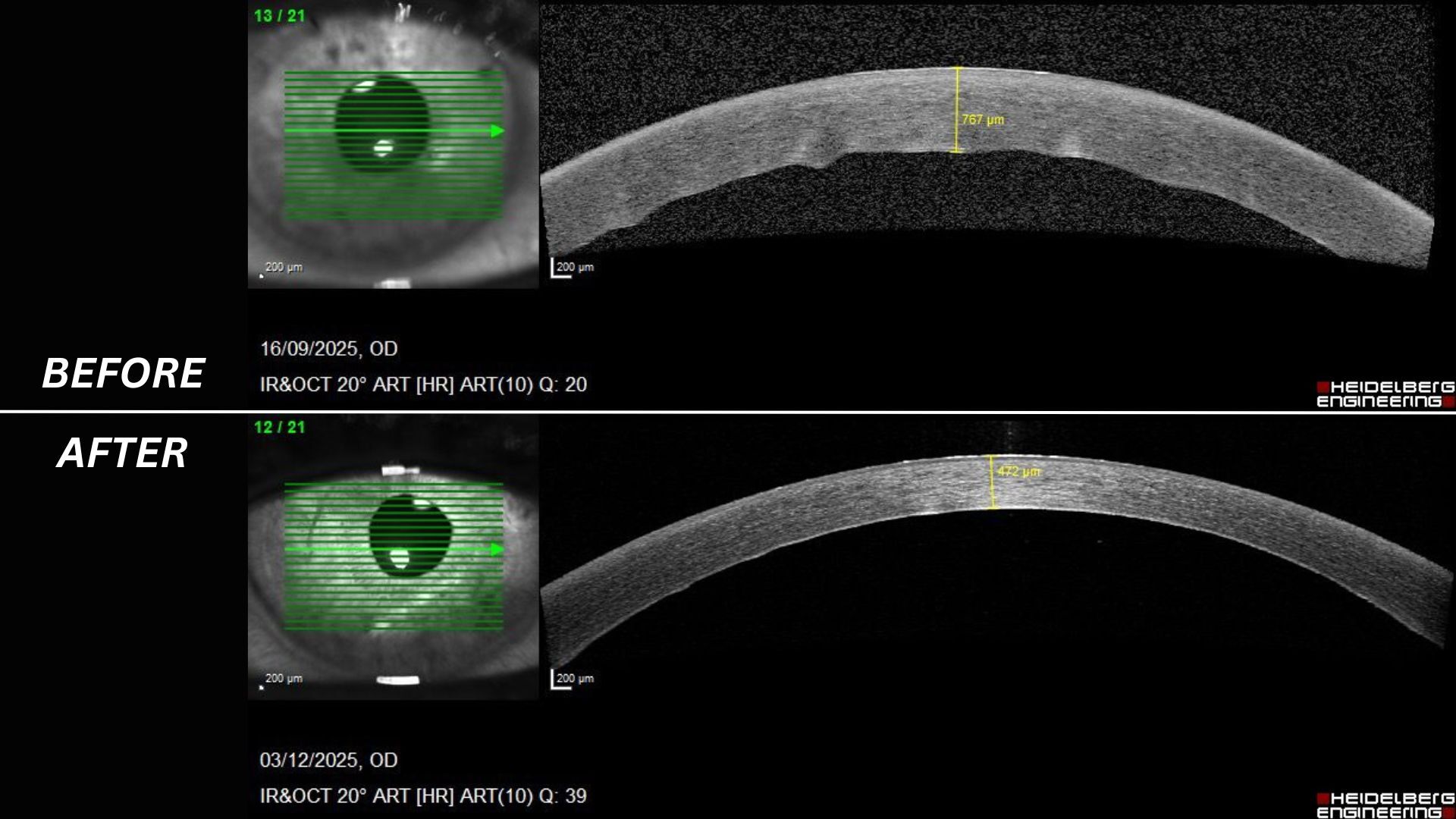 Images courtesy of Precise Bio
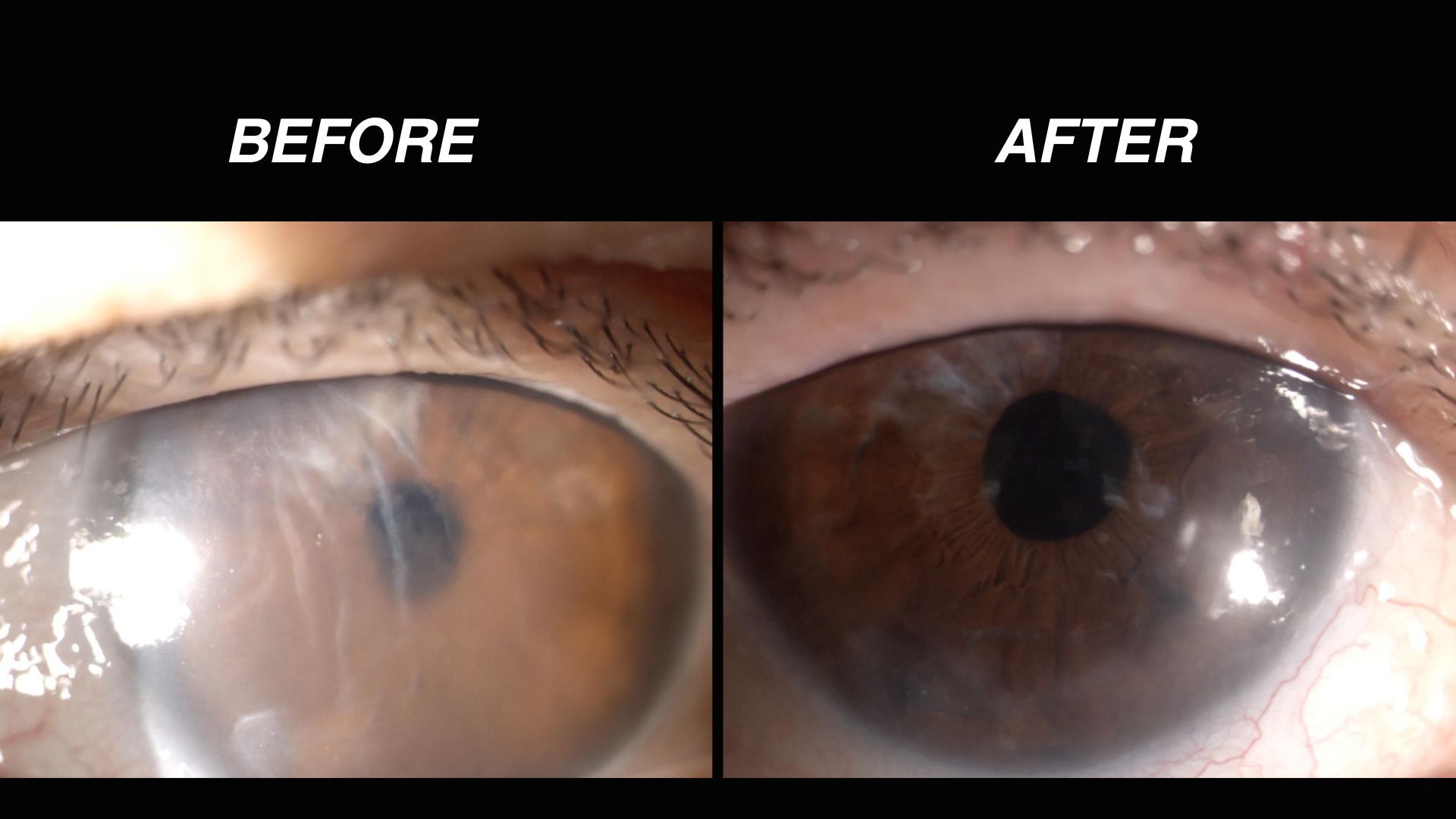 Images courtesy of Precise Bio
NR: How does Precise Bio's approach aim to solve those problems?
AB: At Precise Bio, what we do is fabricate tissues, starting with a donor cornea as our first product. We begin by isolating the cells and developing a proprietary process for proliferating and expanding them. From a single donor tissue, we are able to generate enough cells, a bank of cells, to fabricate over 400 new corneas, which can effectively solve the shortage of donor tissue related to corneal transplants.
There are various other advantages to our tissue as well. For example, it is much easier to transplant than donor tissue. We engineered it with very unique mechanical properties, which shorten operating times and make the procedure much easier for the surgeon.
Another advantage is that, because we print the cells, we can precisely define the cell density of the tissue. While a standard donor tissue provides about 2,000 to 2,500 cells per square millimeter, our tissue has over 4,000 cells per square millimeter. This means the optical outcome of our tissue is expected to be superior to donor tissue.
[Editor's note: Laboratory and animal studies show promising results regarding the ease of the surgery to insert the implants, as well as the cell density within the printed tissue. However, human clinical trials are still ongoing, and therefore, claims that this printed tissue performs better than human donor tissue still need to be confirmed.]
Another important advantage of our tissue, in addition to the availability, is the fact that it is fully tested. We have a production line. And in the production line, the tissue goes to a full quality control test, which also includes viruses and fungus. So when you receive a cornea from our production line versus a donor cornea, ours is fully tested and quality controlled. There's no chance that you're going to have a virus or a fungus that later will affect the tissue in the patient's eye. So these are the advantages of our tissue versus a traditional donor tissue that exists as a treatment today.
[Editor’s note: Traditional donor corneas already undergo rigorous safety screening, including blood testing of the donor for infectious diseases, such as HIV and hepatitis, along with tissue-culture checks of the transplant for fungal and bacterial contamination. Precise Bio's manufacturing model allows for controlled batch testing throughout production and up to transplantation.]
NR: How does the technology work, and how does your printing system ensure that the cornea is uniform and smooth so light can pass through?
AB: The technology is not just the printer; the printer is simply the production tool with which we fabricate the tissue. The technology is a combination of many elements. When we look at natural body tissues, they are composed of cells and the ECM [extracellular matrix], which is the structural material. When we fabricate a tissue, we start from these two components, trying to stay as close as possible to natural tissue by using human cells and natural materials [such as collagen and ECM].
The fabrication process combines the ECM — in most cases, a human collagen-based material — into two layers. One layer is made of collagen, and upon that collagen layer, we print the cells. When I talk about printing cells, think of a standard color printer with red, green and blue cartridges; in our printer, we load human endothelial cells [flat cells that form linings in the body]. We flow these cells through the printhead inside a material called bio-ink, and every laser pulse deposits the cells. This enables us to arrange the cells in the exact anatomical structure they occupy in the body. Post-printing, we manually transfer the tissue from the printer into an incubator, though this final handling will be fully robotic and automated in the future.
NR: The implant can be rolled up, loaded into an injector, and then unrolled inside the eye. What makes the printed material flexible enough to do this without breaking?
AA: Basically, we're using the same material that is present in your very own cornea [collagen] to maintain flexibility, and the printer allows us to deposit the cells precisely where they are needed.
NR: The platform has "single-cell resolution."Why is that important?
AB: The fact that we can arrange cell by cell enables us to mimic the anatomical structure, the exact structure of the tissue of our body. Depositing cells one at a time at very high viability gives us a major advantage in replicating what the body naturally expects.
AA: The best way to explain it is that nature has already figured out the best design through evolution. What this technology does is effectively replicate what nature has already achieved.
Precise Bio is working to develop an alternative approach to corneal transplants, which replace the clear lens at the front of the eyeball. (Image credit: Mark Garlick/science Photo Library via Getty images)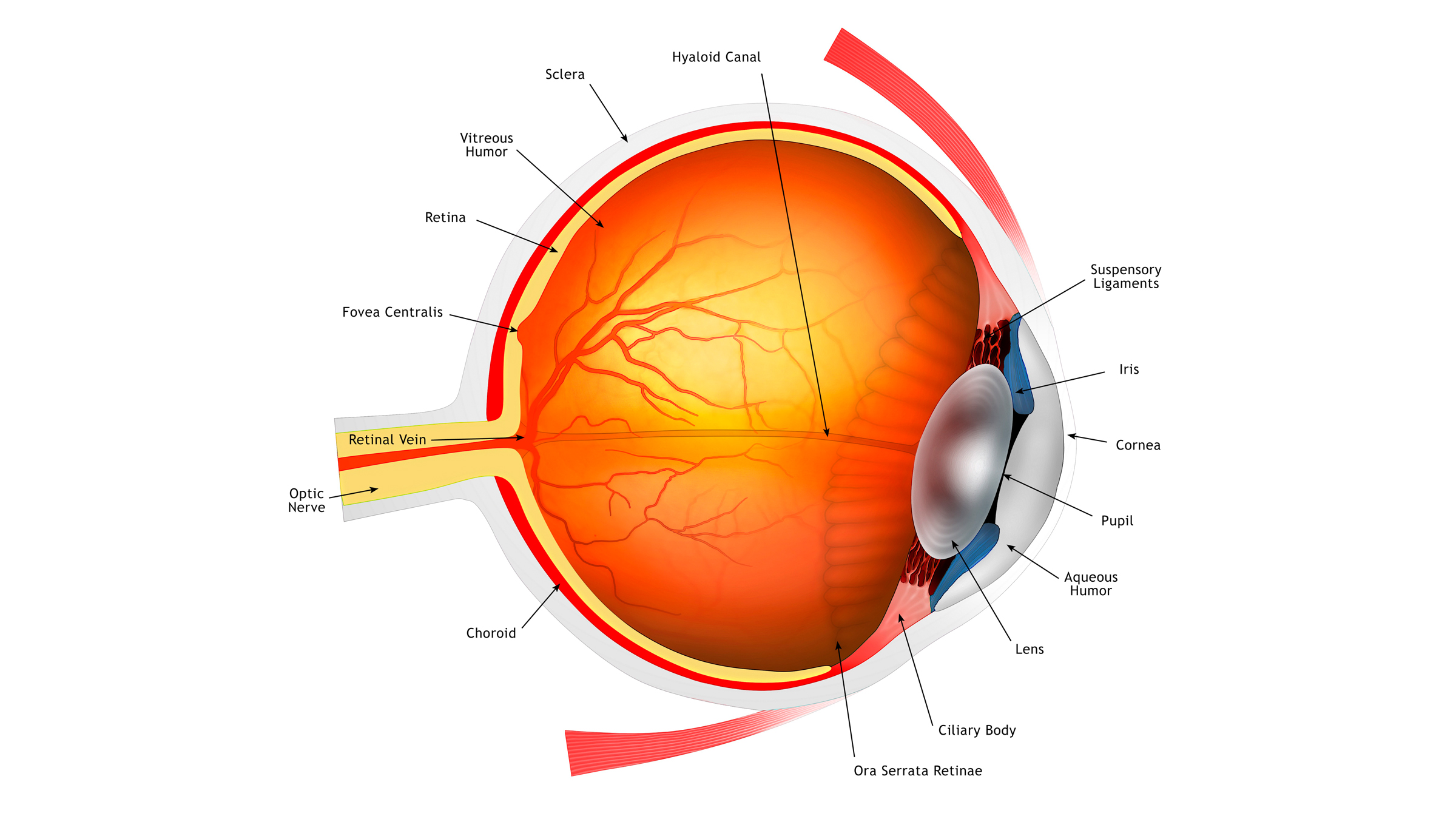
NR: You say these implants could be frozen and shipped worldwide — has that been tested?
AB: While we are currently conducting our Phase I study in Israel, product development has included extensive testing on shipment. We shipped the product from the Middle East to the U.S. and back and transplanted the tissue into animals to verify that it functions after international transit. We had to evaluate shipment times, transport environments and how to maintain tissue properties on a plane.
To assist that, we also developed various technologies related to cryopreservation of the cells and prior preservation of the tissues, which enables us to, at the end of the day, have a supply-on-demand mechanism so we can manufacture anywhere and ship it anywhere in the world for the patient's benefit worldwide. Currently, our tissue is shipped very similar to the tissue that is shipped from a donor tissue, where we have a shelf life of around four or five days.
NR: You've recently conducted the world's first transplant of these new corneas as part of the trial. Could you describe the procedure and how well it worked?
AA: The first patient had been legally blind [in the treated eye] for 14 years.
AB: This specific patient had a non-functioning cornea and could not even count fingers [before the procedure]. This was due to pseudophakic bullous keratopathy, a condition where the cornea swells permanently and forms fluid blisters following cataract surgery.
She is close to nine months now since her transplantation, and she sees well. After several weeks, she was already able to read the menu in a restaurant and subtitles on the television. The most important outcome of this is that now, she can see normally — with her new cornea.
We continue to follow her progress and we will follow up again at the nine-month point and then at the year point. This is essentially the way we do with all of our patients. Now, we already have five patients [who have gotten the new transplants].
AA: [Based on the trial participants they've treated so far] patients recover similarly to those receiving a traditional corneal transplant; visual recovery occurs quickly at first, but continues to improve over time, with initial recovery taking place within days to weeks.
NR: Is there a risk of immune rejection with this kind of implant?
AA: With current techniques using corneas from deceased donors, the cornea is considered "immune privileged," meaning it has features that reduce immune response. Because it lacks blood vessels and lymphatic vessels, the risk of rejection is much lower than with other transplanted organs.
Of course, a small risk always exists with any medical procedure, but even with human bio-printed corneas, the risk is very low and most rejection episodes can be reversed if treated promptly.
AB: There is always some risk of rejection in the eye, but this is taken care of with very mild steroids. In the first weeks, the frequency of the drops is higher, then as the patient moves forward we reduce the need for these steroids.
Severe eye disease, swelling, scarring, and physical trauma to the eye can damage the cornea, causing vision problems that may require a transplant to fix. (Image credit: Francesco Riccardo Iacomino via Getty Images)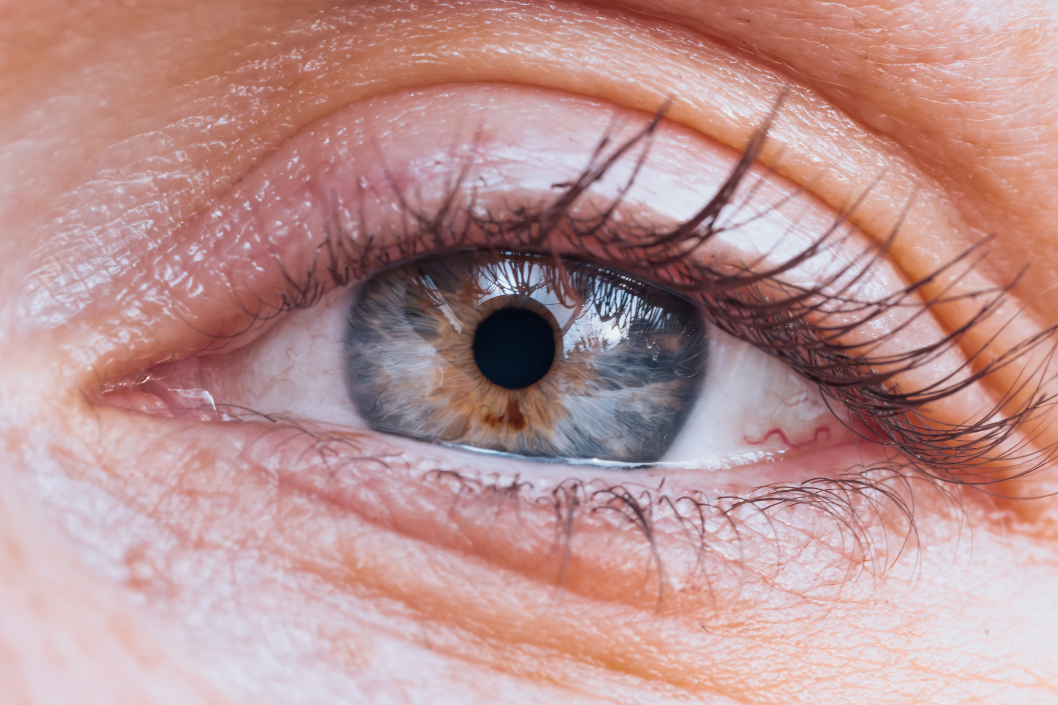
NR: What are the next steps for developing this treatment?
AB: We are in the process of developing longer cryopreservation of the tissue, and we will be completing the Phase I trial in 2026, completing the transplants. And then we will move forward to a more advanced stage. We are planning to transplant an additional 10 patients until the end of the year [as part of the ongoing trial].
After Phase I, we will be submitting an Investigational New Drug [IND]application to the U.S. Food and Drug Administration, where we are targeting to do the more advanced studies. [Approved IND applications grant permission to give a new, unapproved medicine or biological product to clinical trial participants.] The primary focus of this study will be in the U.S., while continuing clinical sites in Europe and Israel, and exploring regions with high demand like India.
In the future, the extended studies that are planned for 2027 will open up to other patients also with other health indications [such as diabetes or high blood pressure].
NR: What timeline do you foresee for getting these new bioprinted corneas fully approved?
AB: We anticipate commercial distribution of our corneas in the U.S. by 2030, followed by expansions into Europe and other regions.
AA: These current studies are still in their very early stages, which is why we are carefully assessing the technology and following patients long-term to ensure the approach can be successfully expanded. As soon as that groundwork is complete, the next phase will move forward in the U.S.
This interview was lightly edited for length and clarity.
This article is for informational purposes only and is not meant to offer medical advice.





 Bengali (Bangladesh) ·
  English (United States) ·